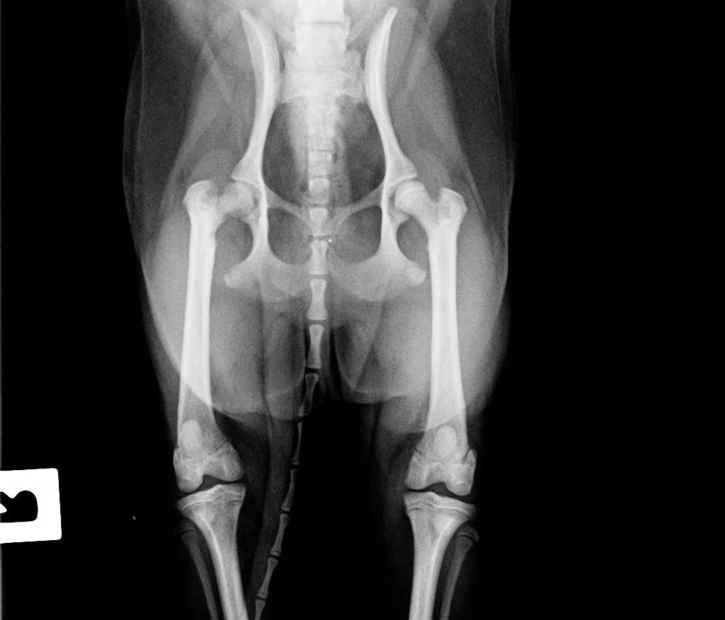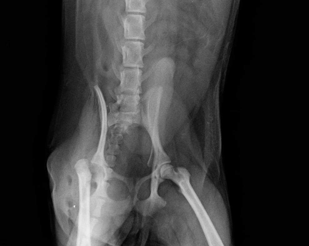
Peach was first seen with an acute onset lameness at the after hours vet clinic. There was no known traumatic incident (ie. falling or tripping).
Peach was found to be painful on examination and was treated with a pain killer. Unfortunately Peach did not get better and kept holding up his little right back leg.
At this stage he came to Mosman Veterinary Hospital and we recommended a radiograph of his pelvis and right hind limb to better assess what might be going on. Take a look at the radiograph below and see if you can spot the difference between the left and the right hip.

It is very subtle! But the right hip has a slightly more ‘lucent’ (see-through) and irregular appearance compared to the left. This is only a slight radiographic change but unfortunately is not good news for the hip joint. This is the classic sign of a disease called Legg-Calve Perthes disease. This is a disease of the hip where the blood supply does not flow effectively up the neck of the femur (thigh bone) to the head of the femur (the ball). This results in effectively the ‘dying off’ of the ball portion of the hip.
Avascular necrosis of the femoral head or Legg-Calve Perthes disease, is a condition that has no known cause. It is common in small and toy breeds and usually only affects one hip. Unfortunately, nothing can be done to save the hip and eventually there is total collapse of the joint which can be painful. The treatment is a surgical procedure called a ‘Femoral Head and Neck Ostectomy’. This procedure removes the ‘ball’ portion of the hip which removes the source of pain. Small dogs undergoing this procedure typically do very well and regain complete function of the leg. The body forms a sort of false joint with fibrous tissue that works extremely well and the patient goes on to live a long and happy life.
Peach had a Femoral Head and Neck Ostectomy at Mosman Vets with Dr Abbie Tipler and Dr Rachele Lowe, which went very well. Peach was a bit painful after surgery and needed some top-up pain killers but otherwise is making a great recovery. The post-operative radiograph looked good and can also be seen below.

Posted on 15 January 2014
Last updated on 12 December 2019
Tagged with: surgery


 Nine Steps to Calm Your Dog in Thunder/Fireworks
